Written by Mark Fretz, Sue Ishaq, and Mira Zimmerman
Light is as necessary to the perfect growth and nutrition of the human frame as are air and food; and, whenever it is deficient, health fails, and disease appears… Artificial is but a very bad substitute for natural light… For health, we cannot have too much light, and, consequently, too many or too large window – The Lancet, 1845
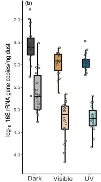
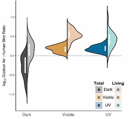
Even with this history of light-based studies, there has been, to our knowledge, no research conducted on the effect of light on the indoor microbial community. The microbial community found indoors is primarily sourced by outdoor air and microbial occupants [22–24]. Many infectious organisms persist indoors for months [25–27], potentially creating a reservoir that may be spread via direct or indirect contact [28]. The presence of microbial cells and cell components can even enhance the allergic reaction of individuals to pet allergens [29]. The building itself, including the materials found indoors and how spaces are used, can determine whether microorganisms survive or perish, remain or are carried away, and whether our methods of cleaning for microbial control is effective [30]. The inclusion of windows, the use of different light filters on glass, shading strategies, and outdoor weather conditions can impact the amount and spectra of light which finds its way indoors, and thus how much daylight the indoor microbial community is exposed to (Figure 1).

Figure 1

Figure 2

Figure 3

Figure 4
To read our publication, click here.
NPR’s coverage of our study is also out, check it out here.
Bibliography
- The Duty on Glass. Lancet 1845, 45:214–216.
- Bazzoni CB: Am. J. Public Health 1914, 4:975–992.
- Ward HM: Lancet 1893, 141:383.
- Downing AMW, Blunt TP: Proc. R. Soc. Lond. 1878, 26:488–500.
- Medeiros AB de A, Enders BC, Lira ALBDC: Esc. Anna Nery 2015, 19:518–524.
- Nightingale F: Notes on Hospitals. Longman, Green, Longman, Roberts, and Green; 1863.
- Hessling M, Spellerberg B, Hoenes K: FEMS Microbiol. Lett. 2017, 364.
- Fonseca MJ, Tavares F: Am. Biol. Teach. 2011, 73:548–552.
- Besaratinia A, Yoon J-I, Schroeder C, et al.: FASEB J. 2011, 25:3079–3091.
- Goldman RP, Travisano M: Evolution 2011, 65:3486–3498.
- Takada A, Matsushita K, Horioka S, et al.: BMC Oral Health 2017, 17:96.
- Dai T, Vrahas MS, Murray CK, Hamblin MR: Expert Rev. Anti. Infect. Ther. 2012, 10:185–195.
- Oppezzo OJ: J. Photochem. Photobiol. B 2012, 115:58–62.
- de Sousa DL, Lima RA, Zanin IC, et al.: PLoS One 2015, 10:e0131941.
- Maclean M, Anderson JG, MacGregor SJ, et al.: J Blood Transfus 2016, 2016:2920514.
- Deng Y, Yao J, Wang X, et al.: PLoS One 2012, 7:e39704.
- Ondrusch N, Kreft J: PLoS One 2011, 6:e16151.
- Patra V, Byrne SN, Wolf P: Front. Microbiol. 2016, 7:1235.
- Prescott SL, Larcombe D-L, Logan AC, et al.: World Allergy Organ. J. 2017, 10:29.
- Sadar JS:. Routledge; 2016.
- Himmelfarb P, Scott A, Thayer PS: Appl. Microbiol. 1970, 19:1013–1014.
- Lax S, Smith DP, Hampton-Marcell J, et al.: Science 2014, 345:1048–1052.
- Prussin AJ 2nd, Marr LC: Microbiome 2015, 3:78.
- Adams RI, Bhangar S, Pasut W, et al.: PLoS One 2015, 10:e0128022.
- Otter JA, French GL: J. Clin. Microbiol. 2009, 47:205–207.
- Kramer A, Schwebke I, Kampf G: BMC Infect. Dis. 2006, 6:130.
- Smith SM, Eng RH, Padberg FT Jr: J. Med. 1996, 27:293–302.
- Hübner N-O, Hübner C, Kramer A, Assadian O: Am. J. Nurs. 2011, 111:30–4; quiz 35–6.
- Herre J, Grönlund H, Brooks H, et al.: J. Immunol. 2013, 191:1529–1535.
- Maclean M, Macgregor SJ, Anderson JG, et al.: J. Photochem. Photobiol. B 2008, 92:180–184.
